Skip to content

Talking to Your Doctor

Each doctor may have their preferred methods of screening, so it’s important to ask questions.
It may seem intimidating at first, but you are your best advocate. 

This valuable discussion guide may help you talk to your doctor about KC, the screening process, and your treatment options

Doctor Discussion Guide

What to expect during an evaluation

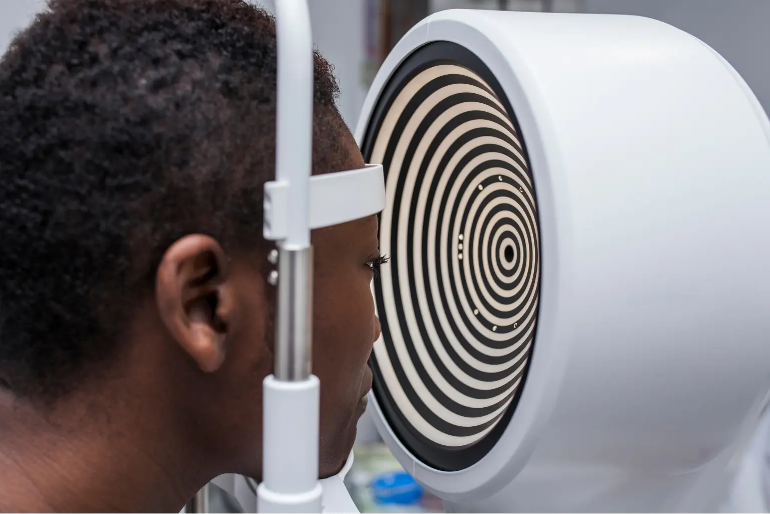
Your doctor will provide a comprehensive eye exam for keratoconus that may include diagnostic tools, such as a corneal topographer, corneal tomographer, and a corneal pachymetry test:

The corneal topographer 

will take a picture of the surface of your eye to detect potential subtle changes of the surface of the eye.

The doctor may also use a corneal tomographer, which looks similar.

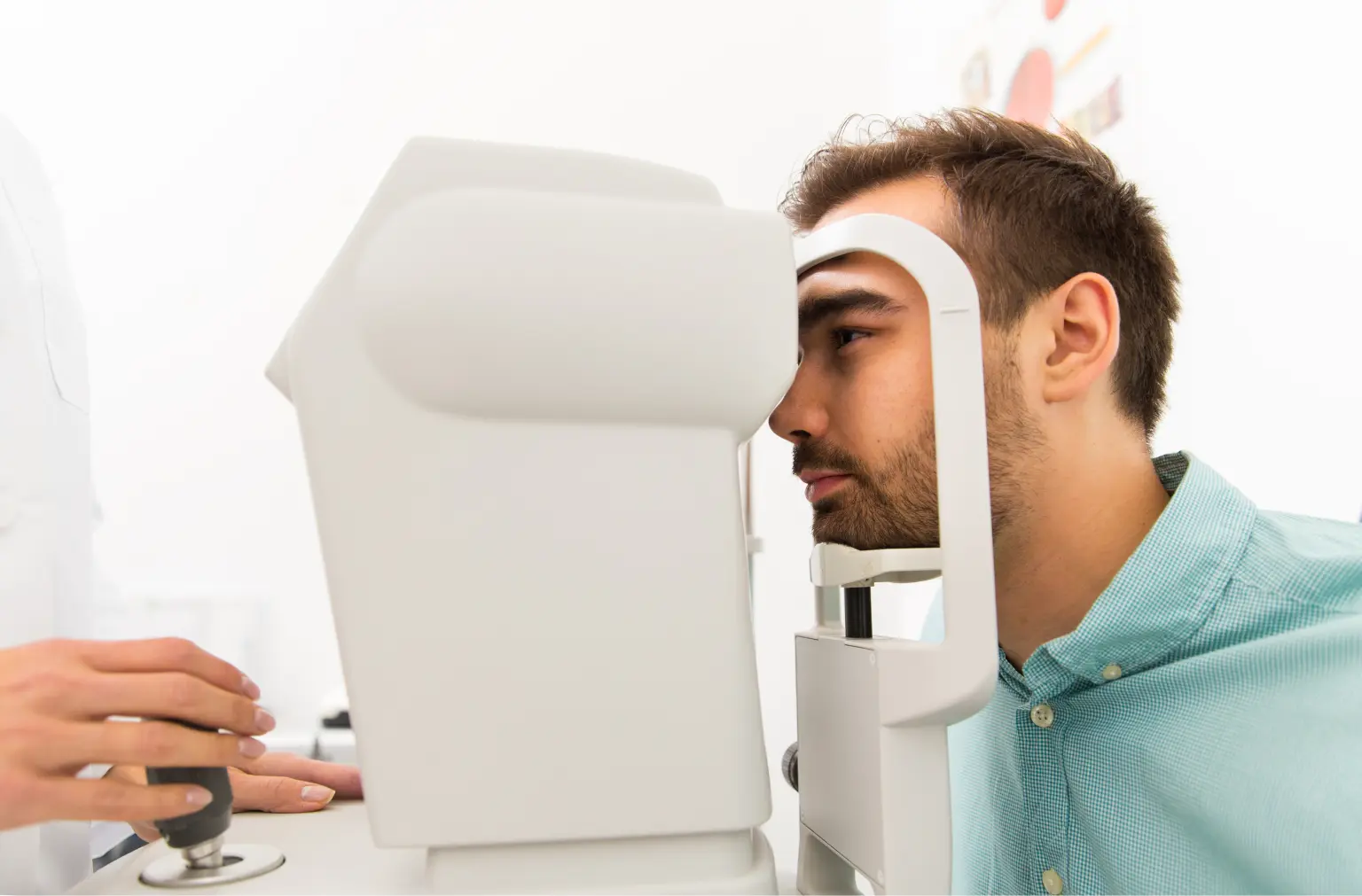
The corneal tomographer 

is an instrument that measures the thickness of the cornea at different locations, especially on the back surface of the cornea. It can be especially valuable because it could detect KC before the cornea starts to form a cone shape.
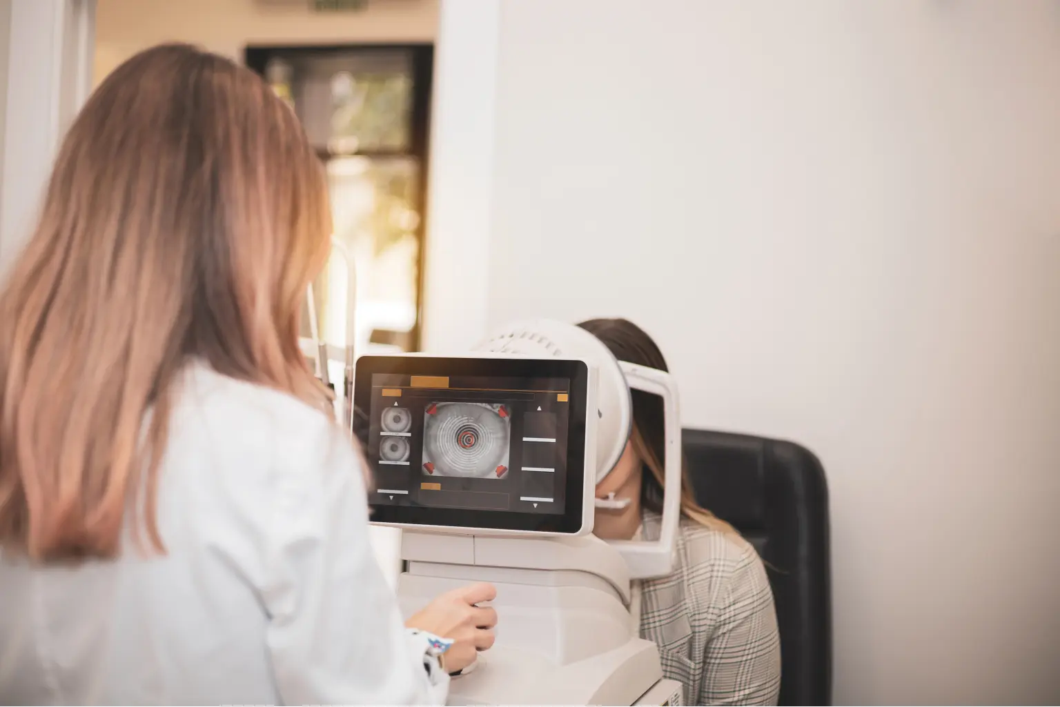
Corneal pachymetry

is a third possible test which measures the thickness of the cornea to show progressive thinning over time.

To get the screening process started, find a doctor near you

Find a Doctor

Living with KC. 
And options for treating it too!

Vision management

Eyeglasses or soft contact lenses Mild or early cases of KC that have not shown progression could benefit from prescription glasses or disposable/reusable contact lenses. These will bend rays of light to focus images on the retina inside of your eye.
Rigid gas permeable contact lenses Rigid gas permeable (RGP) contact lenses are small diameter rigid contact lenses that are placed on the corneal surface. Scleral lenses are large diameter lenses that rest on the sclera (white of the eye) and vault over the cornea. RGP lenses create a smooth uniform surface. Your natural tears or a saline solution fill in the space between the lens and the cornea, “masking” the irregular corneal shape. LEARN MORE ABOUT RGP CONTACT LENSES
Prescription corneal implants Micro-thin prescription corneal implants are designed for the reduction or elimination of nearsightedness and astigmatism, an imperfection in the curve of the eye, in patients with KC. They are thin plastic, semi-circular rings, which are surgically inserted under the surface of the cornea. When inserted into the keratoconic cornea, they flatten the cornea, changing its shape. The placement of this implant may reduce corneal irregularities caused by KC.

Intervention

FDA-approved corneal cross-linking Corneal cross-linking is a minimally invasive outpatient procedure that combines the use of ultra-violet (UV) light and riboflavin (vitamin B2) eye drops. Cross-linking stiffens and strengthens the collagen fibers of the cornea that have been weakened by keratoconus. It may also help KC from getting worse over time. LEARN MORE ABOUT CROSS-LINKING

End-stage surgery

Corneal transplant surgery In KC, when the cornea becomes dangerously thin or when sufficient vision can no longer be achieved by contact lenses due to steepening of the cornea, scarring or lens intolerance, a corneal transplant may be the only option. The surgery is generally performed as an outpatient procedure with either general or local anesthesia. During the surgery, the part of the cornea affected by KC is removed and replaced with a donor cornea. While the surgical transplantation of a new cornea will resolve the basic problem of corneal surface irregularity, eyeglasses or contact lenses are usually needed after the surgery for vision correction.
Stories From the Community
Take the Quiz